Brain Metastases (Brain Mets)
What are Brain Metastases/Metastatic Brain Tumors/Brain Mets?
Brain Metastases (Brain Mets), are secondary tumors that are formed when cancer cells which originate from another part of the body, and spread (metastasize) to the brain.
Certain cancers - specifically lung, breast, and melanoma - are the most likely to spread to the brain. For example, small-cell lung cancer spreads to the brain so often that doctors routinely use preventative radiation therapy to protect it. Other cancers, like prostate or head and neck cancers, rarely spread to the brain. Aside from looking at the exact type and subtype of a person's cancer, it is very difficult to predict who will develop brain metastases.
How does cancer metastasize to the brain?
Cancer metastasize to the brain when it travels through the blood and breaches the blood-brain barrier to enter the central nervous system. As the tumor cells multiply, they invade local tissue, displace surrounding structures, and trigger inflammation and edema (swelling). Although these tumors are most common in high-blood-flow areas, different cell types tend to settle in different regions of the brain.
Metastatic brain tumors frequently lodge at the boundary where gray and white matter meet, heavily influenced by the brain's blood flow patterns. Their typical distribution includes:
-
Cerebral Hemispheres: 80% of metastases
-
Cerebellum: 15% of metastases
-
Basal Ganglia: 3% of metastases
How are Brain Metastases diagnosed?
A proper clinical evaluation requires a thorough history, physical, and neurological assessment, which includes the following:
-
Symptom Screening: Document the duration and intensity of symptoms, specifically noting any headaches, nausea, or blurry vision.
-
Neurological Exam: Assess strength, sensation, reflexes, coordination, balance (proprioception/cerebellar function), cranial nerves, speech, vision, memory, and cognitive thought.
-
Ophthalmic Exam: Inspect the eyes specifically for signs of papilledema.
-
Prognostic Factors: Gather data on the patient's age, performance status, and systemic cancer burden to map out the disease course and guide future therapy.
The imaging and laboratory protocol for suspected brain metastases involves:
-
Initial Screening: A rapid head CT scan for quick evaluation.
-
Definitive Imaging (Gold Standard): A contrast-enhanced, fine-slice brain MRI to pinpoint the exact number of tumors, their anatomical locations, and the severity of accompanying swelling (edema).
-
Baseline Blood Work: A complete blood count, metabolic panel, and liver function tests to assess the patient's overall health.
How are Brain Metastases treated?
The therapeutic pipeline for brain metastases is divided into acute stabilization and definitive, multi-disciplinary interventions.
1. Acute Phase: Edema Management
-
First Line: Corticosteroids (e.g., Dexamethasone).
-
Dosing Example: 10 mg IV bolus, followed by 4 mg IV every 6 hours.
-
Strategy: Rapid taper upon clinical improvement to avoid systemic steroid toxicities.
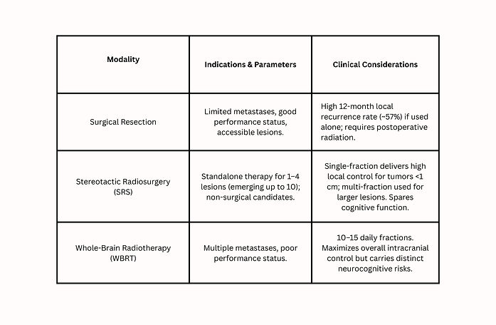
2. Definitive Phase: Treatment Modalities
3. Prognostic Decision Making
-
Post-Op Optimization: Choosing between postoperative WBRT and SRS requires balancing superior tumor control (WBRT) against superior neurocognitive preservation (SRS).
-
Advanced Frailty: In patients with severe functional impairment, WBRT may provide no therapeutic advantage over steroids alone, favoring a palliative, supportive care approach.
Prognosis
How a person is expected to recover or respond to treatment for brain tumors depends on a few important details. Doctors look at the patient's age and overall health, how many tumors there are, and how large they have grown. They also consider where the cancer originally started, whether it has spread to other parts of the body, if the brain tumors are pressing on healthy brain tissue, and how well that specific type of cancer responds to treatments like radiation and chemotherapy.
Potential complications of brain metastases include:
-
Brain Swelling and Pressure: The tumor can push on healthy brain tissue (mass effect), block the natural flow of brain fluid (hydrocephalus), or dangerously shift brain structures out of place (herniation).
-
Nerve and Brain Damage: The cancer can spread into nearby areas, leading to seizures or permanent nerve problems (such as trouble speaking, moving, or remembering).
-
Life-Threatening Risks: If left untreated or if it progresses rapidly, these complications can be fatal.
REFERENCES
Amsbaugh, M. J., & Kim, C. S. (2020, June 30). Brain Metastasis. Nih.gov; StatPearls Publishing. https://www.ncbi.nlm.nih.gov/books/NBK470246/
Bettegowda, C. (n.d.). Metastatic Brain Tumors. John Hopkins Medicine. Retrieved from https://www.hopkinsmedicine.org/health/conditions-and-diseases/metastatic-brain-tumors